One-Year-Old Girl in China Found with Fetus in Her Skull: A Rare Medical Phenomenon

In an astonishing and tragic case, a one-year-old girl in China was discovered to have a fetus trapped inside her skull. This extremely rare condition, known as “fetus in fetu,” affects approximately 1 in 500,000 births. The infant, suffering from severe head swelling and developmental delays, underwent surgery to remove the fetal tissue, but sadly passed away within two weeks due to irreversible brain damage.
Understanding Fetus in Fetu
Fetus in fetu occurs when identical twins, formed from a single fertilized egg, fail to completely separate during early development. As a result, one twin becomes enveloped within the other and can continue to develop partially, forming features like hair, limbs, and fingernails. While this condition often results in the absorbed fetal tissue being located in the abdomen, it has been found in other parts of the body, including the mouth, scrotum, and tailbone. However, its presence in the skull is exceedingly rare and almost always fatal.
A Rare Occurrence in the Skull
According to a report by Xuewei Qin and Xuanling Chen, anesthesiologists from Peking University International Hospital in Beijing, there have only been 18 recorded cases of fetus in fetu within the skull. The report, published in the American Journal of Case Reports, details this particular case where the absorbed twin caused significant complications.
Discovery and Diagnosis
The abnormalities were first detected during a routine check-up at 33 weeks of gestation. Despite this, the baby was born via caesarean section at 37 weeks and appeared healthy enough to go home with her mother. However, as she grew, her head began to swell, and she exhibited developmental delays. At one year old, the child was brought to Peking University International Hospital with severe head swelling and difficulties in standing, speaking, and controlling bodily functions.
Surgical Intervention and Findings
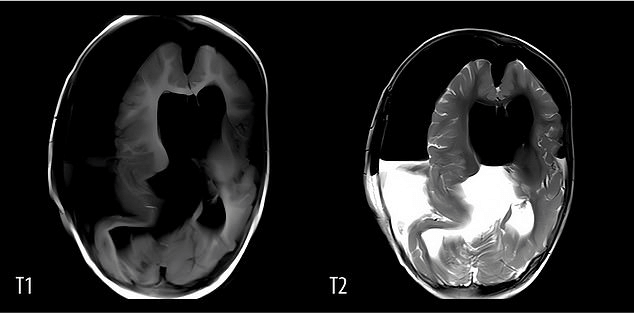
Upon scanning her head, doctors discovered a large mass, about five inches in diameter, inside her skull. The mass contained a white capsule with thick brown fluid and an immature embryo. This embryo had partially developed bones, a spine, and rudimentary features such as a mouth, eyes, hair, forearms, hands, and feet, measuring 18 centimeters in length. The fetus had caused significant compression of the girl’s brain tissue.
Outcome and Reflections
Despite the surgical efforts, the girl’s condition did not improve. She experienced continuous seizures and remained on life support, never regaining consciousness. Twelve days post-surgery, her family made the difficult decision to withdraw life support.
Causes and Theories
The exact causes of fetus in fetu remain unknown. Researchers speculate that environmental factors, genetic anomalies, low temperatures, pesticide exposure during pregnancy, or issues with egg division may contribute to the condition. However, more research is needed to understand the underlying mechanisms fully.
Conclusion
This heartbreaking case underscores the rarity and severity of fetus in fetu, especially when it occurs in the skull. It also highlights the need for further research into the causes and potential prevention of such developmental anomalies. While advancements in prenatal care and medical imaging have improved early detection, cases like this remind us of the complexities and mysteries of human development.
Source: https://www.dailymail.co.uk/health/article-13620831/twin-surgically-removed-skull-China-fetus.html


